
Have you ever heard of an EKG before? For those of you that haven’t, EKG (also referred to as an ECG) stands for an electrocardiogram. An EKG test is a procedure that is used to measure and interpret electrical signals within the heart [2]. You may be wondering how electrical signals are correlated with your heart. Well, each time your heart beats, it sends an electrical signal/current through your heart. Then, the ECG will measure those signals/currents. These electrical signals will help your physician know whether or not your heart is beating at a regular or irregular rate. The latter could be cause for some concern as it can be an indicator of a heart disease [2].
Since an EKG or ECG is focused on the heart and its functioning, it is mainly used to watch out for heart diseases. Some of these heart disorders include heart attacks, heart damage, heart failure, irregular heartbeat, and many more [2]. Along with this, it is also sometimes used during check-ups for patients at a higher risk of heart disease. This higher risk could be a result of family history, diet, lifestyle, or multiple other explanations. Common symptoms to look out for include chest pain, shortness of breath, dizziness, etc. If you are experiencing these types of symptoms, it might be a good idea to get an ECG referral [2]. So what happens during an EKG and how is it conducted? During an EKG, the patient is asked to lay down on a table. After this, a doctor, or another health care provider will place a few electrodes on the patient’s chest, legs, and arms. These electrodes have wires on them that connect them to a computer which is how the electrical signals will be recorded [2]. After this, the physician will analyse the signals and deduce whether anything is irregular or not.
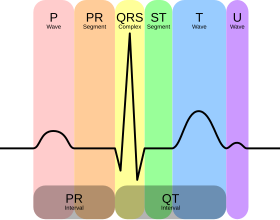
Different parts of an EKG reading correlate to different parts of the heart. If the patient has a normal heartbeat, this will be shown through the EKG by looking at the timing of the upper and lower heart chambers [1]. Now let’s break down the different parts of an EKG. The first wave of an ECG is referred to as the “P wave” and this P wave correlates to the left and right atria (upper chambers of the heart) [1]. The P wave is followed by a flat line which is referred to as the “PR segment” and this segment correlates to when the electrical signals travel to the lower chambers of the heart [1]. There is another wave that follows this flat line, and this wave is referred to as the “QRS” complex. The QRS complex correlates to the right and left ventricles (lower chambers of the heart) [1]. The last wave is referred to as the “T wave” and it correlates to when the ventricles return to a resting state [1].
Overall, EKGs are very important for the prevention of cardiac diseases as they are great indicators of such diseases. EKGs are really beneficial and should be something to keep in mind if feeling any of the above-mentioned symptoms or if you’re a part of a high-risk group.
Author: Priya Amin
References
1. Electrocardiogram (ECG or EKG). http://www.heart.org. (n.d.). Retrieved from https://www.heart.org/en/health-topics/heart-attack/diagnosing-a-heart-attack/electrocardiogram-ecg-or-ekg
2. U.S. National Library of Medicine. (2020, December 10). Electrocardiogram: Medlineplus medical test. MedlinePlus. Retrieved from https://medlineplus.gov/labtests/electrocardiogram/#:~:text=An%20electrocardiogram%20(EKG)%20test%20is,a%20normal%20rate%20and%20strength.

Leave a comment